en.wikipedia.org
Acanthocephala — Wikipedia
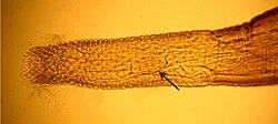
The phylum of thorny-headed worms to which Intraproboscis belongs — obligate intestinal parasites found in vertebrates worldwide.
Wikipedia's Featured Article for May 22, 2026 is Intraproboscis, a genus of thorny-headed worm first described in 2021 after a five-year-old black-bellied pangolin died from intestinal perforation in the Central African Republic. Its sole species, I. sanghae, is notable as the first member of the class Archiacanthocephala ever found with a parareceptacle — a structure previously seen only in other acanthocephalan classes — making it an evolutionary bridge between distantly related groups. Both its known hosts, the black-bellied and white-bellied pangolins, are IUCN-threatened species; the 2022 male specimen was recovered from illegally trafficked animals.

The phylum of thorny-headed worms to which Intraproboscis belongs — obligate intestinal parasites found in vertebrates worldwide.
The original host species of Intraproboscis sanghae — a Vulnerable African pangolin found across central and west Africa.
Wikipedia's Featured Article for May 22, 2026: the full article on Intraproboscis sanghae, the newly described acanthocephalan parasite of African pangolins — taxonomy, morphology, life cycle, hosts, and conservation context.
このコンテンツについて、さらに観点や背景を補足しましょう。